MRI (Magnetic Resonance Imaging)About the MRI
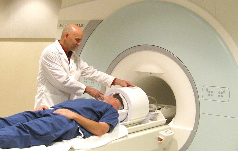
The MRI is a medical tool used to diagnose diseases or injuries in the human body. It consists of a large tube with a table in the middle, to allow the patient to slide in. This machines uses a large magnet, radio waves, and a computer to produce a 3D image for doctors to distinguish any abnormality. Unlike an x-ray, a MRI does not use ionizing radiation, therefore it is considered one of the safest diagnosing equipments. However, the magnetic waves can damage metal devices in the body such as a pacemaker. |
|
About the Procedure
Click on the button below for a video on the procedure.
- The Technologist will go over the MRI screening form with the patient, explain the procedure and answer any questions that the patient may have.
- Patients will be offered headphones to help reduce the loud humming and thumping sounds which are produced during the scanner's operation. The patient is then be escorted into the scan room.
- The scan room has a large doughnut-shaped magnet with a padded table that moves the person into the centre of the machine once the patient is positioned. Whether or not the patient enteres the machine head first or feet first, or how far in the patient goes, is determined by the type of exam performed. In some cases a special piece of equipment called a surface coil, which is like a radio antenna, is placed on or wrapped around the body part that is being imaged.
- The scanner is equipped with an emergency assistance alarm button, a two-way intercom for communication with the Technologist .
- It is very important that the patient remains relaxed and try not to move during the scan. Even very slight movement of the part being scanned can cause distorted images that will have to be repeated. For some exams patients may be asked to hold their breath for 10-30 seconds as the pictures are taken
Click on the button below for a video on the procedure.
|
Uses of MRI
A MRI is useful to diagnose cancer in areas such as the brain, breasts, and soft tissues, as well as seeing how far the cancer has progressed to the rest of the body. The results can provide a better understanding of a change in shape of an organ due to an injury or disease, or the shape, size, and location of the tumour. Results of MRI Can Show:
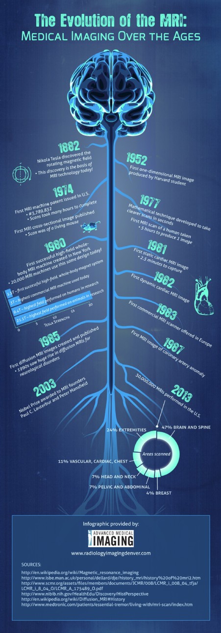
History of the MRI 1946- Felix Bloch and Edward Purcel discovered magnetic resonance. This technology was used for chemical and physical analysis until 1970s. 1952- Felix Bloch and Edward Purcell are awarded the Nobel Prize. 1971- Dr. Raymond Damadian observed that in the presence of nuclear magnetic resonance, tumours found in mice displayed different signals compared with normal tissues. 1977- The first human body scan was produced, taking a total of five hours. The first MRI scanner is currently in the Smithsonian Institute. 2003- Paul C. Lauterbur and Peter Mansfield are awarded the Nobel Prize for discovering the use of MRI as a diagnosing tool. *The image on the right depicts the evolution of the mechanics of MRI dating back to the discovery of rotating magnetic fields. |
|
Created By:
E.C and E.B for Mr. Oh
